UmiCS
Robotic Cardiac Surgery
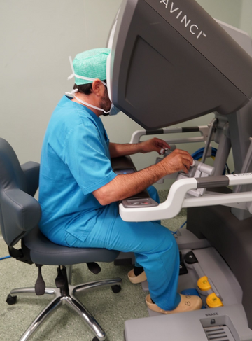
Robotic Cardiac Surgery is a minimally invasive technique that uses a surgical robot controlled by a surgeon to perform complex cardiac procedures.
Book an Appointment
For any clarification, you can contact our Customer Service at 911 501 808.

WHAT IS THE 3D CHEST WALL STUDY
When the patient comes to see the thoracic surgeon (a specialist in chest wall deformities), a 3D scan of the chest wall is carried out to assess the degree of the deformity.
Description of the procedure
Before establishing a suitable treatment plan for Pectus Excavatum, the severity of the deformity must be stratified. Your specialist doctor will carry out complementary tests to confirm the diagnosis and screen for associated pathology. In addition, other complementary diagnostic tests may be carried out, including pulmonary function tests, chest X-ray, electrocardiogram (ECG) and echocardiogram. As the morphology of the deformity varies greatly between patients, it is recommended that a preoperative thoracic computed tomography (CT) scan be carried out to document the morphology, dimensions and severity of the depression (Haller index / depression index) of the thorax.
Nowadays we perform a 3D chest wall study with a computer without radiation to the patient. This study then allows us to order a non-surgical treatment system to treat pectus excavatum (Vacuum Bell) or pectus carinatum (orthostatic vest).
Duration
> 10 min
Anesthesia
> -
Hospitalization
> -
Medical leave
> -
Recovery
> -
Inhibition of physical activity
> -
Learn more
about this
procedure
Make an appointment to clarify any doubts or call us through our contacts