UmiCS
Robotic Cardiac Surgery
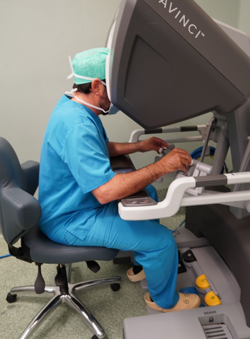
Robotic Cardiac Surgery is a minimally invasive technique that uses a surgical robot controlled by a surgeon to perform complex cardiac procedures.

Book an Appointment
For any clarification, you can contact our Customer Service at 911 501 808.

What are chest wall deformities?
Congenital deformities of the chest wall are a very common pathology in our population and one that is becoming more and more important than in years gone by. Technological and digital developments have allowed young people to research and find out more about this pathology, contributing to a greater understanding of the deformity and its treatment.
These deformities are generally divided into five categories: Pectus Excavatum, or hollowed chest, Pectus Carinatum, or keeled chest, Poland's syndrome, sternal defects and thoracic deformities associated with systemic skeletal diseases.
Among the group of deformities, the most common is Pectus Excavatum, with an incidence in the general population of 1:500, four times more frequent in males, followed by Pectus Carinatum, with an incidence four times lower. In addition, there are also mixed alterations combining characteristics of both deformities mentioned, which can be asymmetrical (more pronounced in one half of the thorax than the other) or unilateral (only on one side).
They usually appear in the anterior part of the thorax, where the ossified structure is extended by cartilage that connects to the sternum. The arrangement and distribution of collagen in the costal cartilage is altered and develops abnormally when subjected to tension, compression and flexion, and it is this deep growth that causes the anterior wall (including the sternum) to sink or the wall to protrude, Pectus Excavatum and Pectus Carinatum, respectively.
In the first few years of life, deformities may begin to appear, but in the vast majority of cases they are detected during adolescence due to faster growth.
These bone deformities of the chest wall are sometimes associated with breast hypoplasia (incomplete development of one breast) on the same side as the deformity and partial absence of some ribs.
This group of deformities includes all types of chest wall deformities except those of the thoracic spine, the latter of which are usually associated with other spinal alterations, such as scoliosis and lordosis, which are treated by Orthopaedics.
Procedures
Pectus Escavatum
Non-invasive treatment with Vacuum Bell
In younger patients, Pectus excavatum can be treated with a suction cup device, Vacuum Bell.
3D chest wall study
When the patient comes to see the thoracic surgeon (a specialist in chest wall deformities), a 3D scan of the chest wall is carried out to assess the degree of the deformity.
Surgery with suprasternal silicone prosthesis
It is one of the minimally invasive solutions for the treatment of Pectus Excavatum and Poland's Syndrome.
Nuss endoscopic surgery
Technological developments have allowed the endoscopic technique used to treat pectus excavatum to become less invasive - Nuss surgery.
Pectus Carinatum
Pectus Carinatum Surgery
The surgery to correct Pectus Carinatum through minimally invasive approaches uses the Abramson Technique, which has been adopted for years in major international Thoracic Surgery reference centers and has a high success rate.
Non-invasive treatment with an orthostatic vest
Once the 3D study has been carried out, the first step can be to opt for non-surgical treatment by fitting each patient with a custom-made orthostatic vest. This vest will compress the sternum to position it in the normal position.
3D chest wall study
When the patient comes to see the thoracic surgeon (a specialist in chest wall deformities), a 3D scan of the chest wall is carried out to assess the degree of the deformity.